The main difference between light and electron microscopes is the radiation used to form an image. Light or optical microscopes are characterized by light as a source of illumination and optical lenses as magnifying objects while in electron microscopes these functions are performed by electrons and electromagnetic lenses respectively.
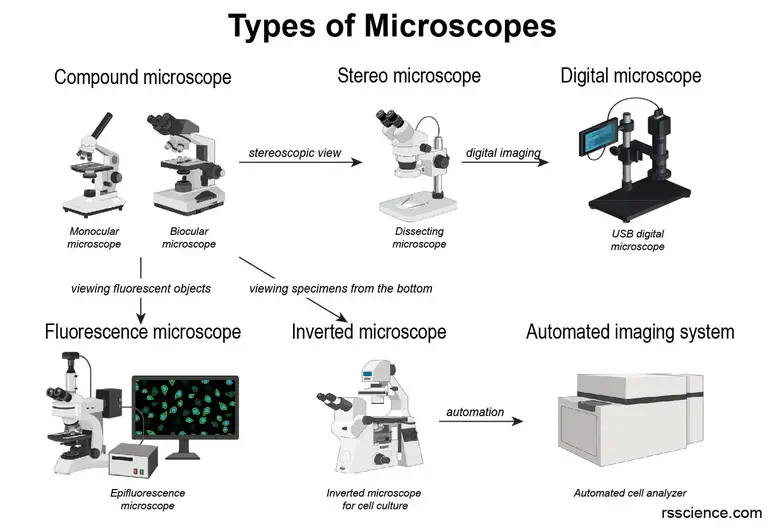
Different Types Of Microscopes Light Microscope Electron Microscope Scanning Probe Microscope Rs Science
The light microscope or optical microscope is a microscope that uses visible light and a system of lenses to magnify images.
. No risk of radiation leakage. It uses a beam of electrons rather than visible light as the light source to make objects larger and present a more detailed picture. It has become an indispensable tool in microbiology and quality control and assurance.
The resolution is well-matched to the sizes of subcellular structures a diverse range of available fluorescent probes makes it possible to mark proteins organelles and other. While a light electron simply uses light to magnify an object an electron microscope uses electrons that are then passed through a specimen. If you think of an electromagnetic spectrum visible light is in the middle.
Before the invention of electric bulbs microscopists were limited in their choice of suitable sources for microscope illumination. The electron microscope was invented in 1931 by the physicist Ernst Ruska and engineer Max Knoll. 20x Greater Depth-of-Field Multi-Angle Observation Advanced 2D3D Measurement.
It is also used in metallurgy. Typically the condenser focuses the image of the light source directly onto the. It is a special type of microscope with a high resolution of images as the images can be magnified in nanometers.
Specimen preparation takes about a few minutes or an hour. Electron microscopes allow for higher magnification in comparison to a light microscope thus allowing for visualization of cell internal structures. Ad Improve observation and analysis capabilities with our 4K Digital Microscope.
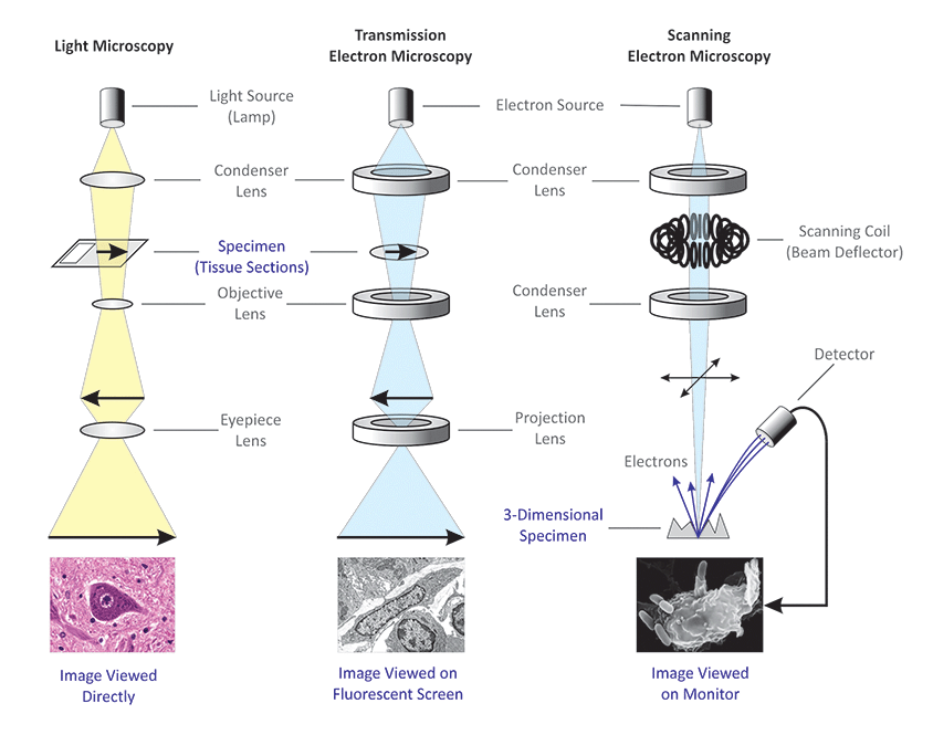
When examined with the electron microscope the terminal bars were resolved to be junctional. Light microscopy has several features that make it ideally suited for imaging biology in living cells. The light microscope left the transmission electron microscope TEM center and the scanning electron microscope SEM right.
There are two types of electron microscopes. These days there are many complex designs of them which have been developed with the aim of improving resolution and sample contrast. Optical and electron microscopes are often used effectively despite little knowledge of the relevant theory or even of how a particular type of microscope functions.
Comparing image formation in light and electron microscopes. As the wavelength of an electron can be up to 100000 times shorter than that of visible light photons electron microscopes have a higher resolving power than light microscopes and can reveal the structure of smaller objects. Specimen preparation takes several days.
Terminal bars as viewed by light microscopy are sites of apparent attachment of epithelial cells that have been shown to be structures that are continuous around the circumference of the entire cellTerminal bars occupy restricted regions of the cell located in the vicinity of its apex. These lamps are controlled by external power supplies that are designed to meet the electrical requirements of first igniting the lamp then providing the correct current to maintain constant illumination. In epipolarization microscopy the light passes through the objective before it strikes the specimen and then the reflected light is captured by the same objective lens.
During daylight hours they could point their microscopes or substage reflector mirrors towards the sky and use the clouds as a crude diffusion screen to spread illumination evenly across the entire field of view. The resolving power of the electron microscope is far greater than that of the light microscopes. Using visible light as a radiation has several limitations which the electron microscope lessens.
An electron microscope is a microscope that uses a beam of accelerated electrons as a source of illumination. In a modern microscope it consists of a light source such as an electric lamp or a light-emitting diode and a lens system forming the condenser. 42 Light Microscopy Microscopes using light can be categorized into following types.
Covalent Delivers Expert Analysis for Advanced Materials Device Innovation. Uses light approx 400-700 nm as an illuminating source. The condenser is placed below the stage and concentrates the light providing bright uniform illumination in the region of the object under observation.
Ad Support Client Success with a Broad Range of Industry Expertise and Advanced Instruments. Due to the use of a shorter wavelength of electrons better resolution is obtained. Light microscopy vs electron microscopy.
There are numerous light sources available to illuminate microscopes both for routine observation and critical photomicrography. A light microscope is an optical microscope which uses a ray of light to view the image where a condenser collects the light and diverges it to the specimen. Polarized light microscopes can be used in the reflected light or epi-illumination mode.
Risk of radiation leakage. Electron microscopes use a beam of electrons opposed to visible light for magnification. Arc Lamps - Mercury vapor xenon and zirconium arc lamps are also useful sources of illumination for specialized forms of microscopy.
LM which uses visible light as a source of illumination and optical glass lenses to magnify specimens in the range between approximately 10 to 1000 times. Thin sectioning immuno-labeling negative staining to answer specific questions. Light microscopy typically uses wavelengths of light in the visible spectrum which inherently limits it spatial resolution due to the Rayleigh criterion to approximately half the wavelength used approximately 200 nm at best.
It has comparatively a low-resolution and magnification power than the electron microscope which is about 02 µm and 500 1000 X respectively. Electron microscopy is used in conjunction with a variety of ancillary techniques eg. However light microscopes are much more practical in general use.
Electron microscope uses the electron beam as a source of illumination and is used to examine structures too small to be resolved with light microscopes. Many years ago carbon arc lights or zirconium bulbs were used to achieve high levels of illumination but these antique sources are seldom seen today because the lamps reduced the quality and homogeny of light reaching the sample. When optimized illumination of the specimen should be bright glare-free and evenly dispersed in the field of view.
Light microscopy is a key tool in modern cell biology. Uses electron beams approx 1 nm as an illuminating source. A most common light source because of its low cost and long life is the 50 or 100 watt tungsten halogen lamp as illustrated at the base of.
Definition of Electron Microscope. An electron microscope is defined as the type of microscope in which the source of illumination is the beam of accelerated electrons. Lower magnification than an electron microscope.
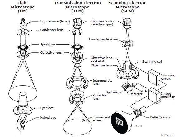
The light and electron in the names refer to the radiation being used. It is a special type of microscope having a high resolution of images able to magnify objects in nanometres which are formed by controlled use of electrons in a vacuum captured on a phosphorescent screen. Figure 1 shows schematic cross-sections of imaging modes for three types of microscopes regularly used for materials characterization.
Light microscopes use visible light which passes and bends through the lens system. Different Types of Light Microscopy. An electron microscope is a microscope that uses a beam of accelerated electrons as a source of illumination.
Increasingly light emitting diodes LEDs are used as a source for microscope illumination in transmitted light as well as fluorescence. The high resolution of EM images results from the use of electrons which have very short wavelengths as the source of illuminating radiation.
Microscopy For Materials Characterization Illuminating Structures With Light And Electrons American Laboratory

Light Microscope Vs Electron Microscope Accelerating Microscopy
Differences Between Light Microscope And Electron Microscope

Transmission Electron Microscope Tem Introduction To Jeol Products Jeol Ltd


0 Comments